Sir,
Among the myriad advantages of musculoskeletal ultrasound (MSUS), its capability to quantify the dimensions of several structures in the musculoskeletal system is noteworthy (1, 2). On the other hand, bearing in mind the most important disadvantage of MSUS (i.e. user-dependency), such measurements sometimes provoke scepticism. We have shown recently that novice sonographers tend to measure small structures smaller and larger ones larger, and that the difference between their measurements and those of an expert depends on experience (3). To investigate this further, in this small study we have tried to illustrate in detail how such measurements changed in the very early period of learning in MSUS.
After an intense two-day course on shoulder ultrasound for novice physiatrists, one novice (LT) and one expert sonographer, with more than 10 years of MSUS experience (LÖ), measured 4 parameters on the same model for 10 consecutive days. Biceps tendon short/long axes, supraspinatus tendon thickness and acromio-humeral distance were quantified in the non-dominant shoulder of a healthy colleague. Statistical Package for Social Sciences (version 16.0, SPSS Inc, Chicago, IL) was used for statistical analysis.
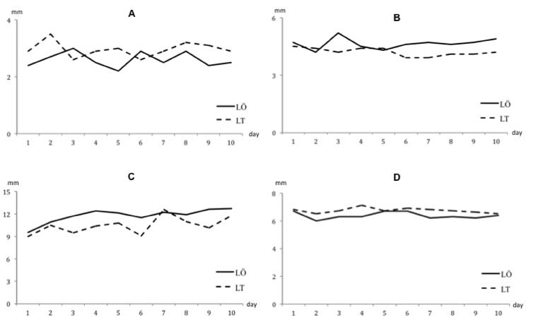
Fig. 1 shows the comparison between the measurements of the novice vs the expert. The two sonographers’ measurements were statistically different (all p < 0.05) and there were no correlations between them (all p > 0.05). On the other hand, intraclass correlation coefficients were high for both expert’s and novice’s measurements (0.98 and 0.96, respectively).
Fig. 1. Comparison between novice (LT) and expert (LÖ) sonographers measurements concerning (A) biceps short axis, (B) biceps long axis, (C) acromio-humeral distance, and (D) supraspinatus thickness.
In the light of our findings, we conclude that, in the early period of MSUS education, the measurements of novice sonographers are “consistently” different from those of experts. Our results comprise only a snapshot regarding an individual scenario. However, it seems that there is crescendo of use of MSUS in the realm of Physical and Rehabilitation Medicine (4) (which should be supported as much as possible), and once physicians start learning MSUS, they naturally start quantifying structures. In this regard, we suggest that such data should be interpreted attentively and that novice sonographers should avoid reporting “rigid” numbers before they gain sufficient experience. Lastly, one way to overcome this impediment may be to use repeat measurements and to consider their mean values for possible evaluation/analysis.
References
Submitted May 30, 2011; accepted July 7, 2011
Levent Tekin, MD1, Murat Kara, MD2, Türker Türker, MD3 and Levent Özçakar, MD4*
From the 1Department of Physical Medicine And Rehabilitation, Gülhane Military Medical Academy, Haydarpaşa Training Hospital, Istanbul, 2Department of Physical Medicine and Rehabilitation, Ankara Physical Medicine and Rehabilitation Education and Research Hospital, 3Department of Public Health, Gülhane Military Medical Academy and 4Department of Physical Medicine And Rehabilitation, Hacettepe University Medical School, Ankara, Turkey. *E-mail: lozcakar@yahoo.com

