Keiji Hashimoto, MD, PhD1 and Masahiro Abo, MD, PhD2
From the 1Division of Rehabilitation Medicine, National Center for Child Health and Development and 2Department of Rehabilitation Medicine, The Jikei University School of Medicine, Tokyo, Japan
Keiji Hashimoto, MD, PhD1 and Masahiro Abo, MD, PhD2
From the 1Division of Rehabilitation Medicine, National Center for Child Health and Development and 2Department of Rehabilitation Medicine, The Jikei University School of Medicine, Tokyo, Japan
OBJECTIVE: The diagnosis and management of mild traumatic brain injury continues to be a subject of debate, with varying opinions regarding the extent to which organically based impairments vs the impact of other stressors cause ongoing disability. The aim of this study was to elucidate the possible abnormalities in benzodiazepine receptor uptake in the brains of patients with mild traumatic brain injury. Nine unmedicated patients with mild traumatic brain injury were investigated using 123I-iomazenil single photon emission computerized tomography (SPECT).
DESIGN: A descriptive study comparing patients after mild traumatic brain injury with matched control subjects.
SUBJECTS: Nine patients with mild traumatic brain injury and 5 controls.
METHODS: The SPECT scan was taken 180 min after injection of tracer.
RESULTS AND CONCLUSION: All 9 patients had a significant increase (> 2 standard deviations higher than the mean of controls) in benzodiazepine receptor uptake in the prefrontal cortex and significantly higher frontal association cortex-to-average global brain activity ratios than in controls. This SPECT study demonstrated focally altered benzodiazepine receptor uptake in the prefrontal cortices in patients with mild traumatic brain injury.
Key words: mild traumatic brain injury, MTBI, single photon emission tomography, SPECT, neuropsychological dysfunction.
J Rehabil Med 2009; 41: 661–665
Correspondence address: Keiji Hashimoto, Division of Rehabilitation Medicine, National Center for Child Health and Development, 2-10-1 Okura, Setagaya, Tokyo, 157-8535, Japan. E-mail: hashimoto-k@ncchd.go.jp
Submitted September 30, 2008; accepted April 1, 2009
INTRODUCTION
Trauma patients frequently report neurological (e.g. headache, dizziness) and psychological (e.g. fatigue, anxiety) symptoms following acute mild traumatic brain injury (MTBI). Acute neuropsychological difficulties with attention, information processing and memory generally resolve within 1–3 months (1). On the other hand, a considerable number of patients continue to report 3 or more symptoms after 3 months. The aetiology of these persistent symptoms remains controversial, partly because the symptoms are not specific to MTBI, being found in other clinical conditions and in the normal population (2).
In order to address the public health problem of MTBI and to help clinicians who treat this disorder, there is a need to identify the best scientific evidence in support of prevention, diagnosis, prognosis and treatment. In addition, gaps in knowledge should be identified, and these areas studied in order to enhance the body of existing scientific knowledge (3). Establishing a diagnostic method to detect abnormalities in the brain of patients with MTBI is an important step in this effort.
Davolos et al. (4) reviewed the utility of single-photon emission computerized tomography (SPECT) as a diagnostic tool in MTBI. The results of this review suggested that SPECT may be a useful tool in the detection of MTBI and in planning treatment. Bonne et al. (5) reported regions of hypoperfusion in frontal, pre-frontal and temporal cortices and sub-cortical structures in MTBI, as shown by Tc-99m-HMPAO brain SPECT imaging. Gowda et al. (6) reported that Tc99m-ECD SPECT can be used as a complementary technique along with computed tomography (CT) in the initial evaluation of patients with MTBI. In addition, it would be particularly useful in patients with post-concussional syndrome, loss of consciousness, or post-traumatic amnesia and a normal CT scan. However, in clinical situations, there are limitations in diagnosing MTBI by conventional magnetic resonance imaging (MRI) and SPECT because of the wide variety of symptoms and neuropsychological dysfunction. Sometimes we cannot explain the neuropsychological problems solely through subtle abnormal findings by MRI and SPECT.
The objective of this study was to detect the organically-based impairment specific to MTBI by using 123I-iomazenil SPECT.
PATIENTS AND METHODS
Patients
We strictly followed the diagnostic criteria of the WHO collaborating centre for the neurotrauma task force on MTBI (7). The task force recommended the following operational definition of MTBI: MTBI is an acute brain injury resulting from mechanical energy to the head from external physical forces. Operational criteria for clinical identification include: (i) presence of one or more manifestations that include confusion or disorientation, loss of consciousness for 30 min or less, post-traumatic amnesia for less than 24 h, and/or other transient neurological abnormalities, such as focal signs, seizure, and an intracranial lesion not requiring surgery; and (ii) a Glasgow Coma Scale score of 13–15, 30 min post-injury or later upon presentation for healthcare. These manifestations of MTBI must not be due to drugs, alcohol, or medication; caused by other injuries or treatment for such other injuries (e.g. systemic injuries, facial injuries or intubation); due to other problems (e.g. psychological trauma, a language barrier or coexisting medical condition); or the result of a penetrating craniocerebral injury.
Nine patients (6 males, 3 females) newly diagnosed with MTBI suffering from any symptoms (headache, dizziness, fatigue, poor concentration, memory problems and anxiety) were investigated using SPECT scanning with 123I-iomazenil as the tracer. Mean age (standard deviation (SD)) was 40.4 (SD 12.1) years (range 22–57 years), and they were not being medicated at the time of the SPECT studies. Five age- and sex-matched and unmedicated controls (4 males, 1 female; mean age 37.6 (SD 18.1) years, range 18–58 years) with no history of MTBI or any neuropsychological dysfunction participated in this study. To assess activities of daily living and neuropsychological dysfunction of MTBI patients, the Functional Independence Measure (FIMTM and the Wechsler Adult Intelligence Scale Revised (WAIS-R) were administered. Informed consent was obtained from all participants after a full explanation of the study procedure.
SPECT
Imaging technique. 123I-IMP (N-isopropyl-4-iodoamphetamine (123I) hydrochloride) SPECT and 123I-IMZ iomazenil (123I)) SPECT images were obtained by a 30-min acquisition beginning at 20 min after the administration of 222 MBq of IMP and 3 h after the administration of 222 MBq of IMZ. The SPECT was performed with a triple-head rotating camera (Siemens, Munich, Germany, OPEN Multi SPECT3) under standard resting conditions (eyes closed) in all study subjects. The distance of the collimators from the centre of rotation was kept constant at 13.8 cm. A fan beam collimator and a 128 × 128 acquisition matrix were used for data acquisition. Images were reconstructed with a Butterworth filter (123I-IMP: cut-off frequency 0.27 nyquist, order 5; 123I-IMZ: cut-off frequency 0.25 nyquist, order (8). Pixel size in the reconstructed images was 2.46 mm/pixel. Attenuation was corrected according to Chang’s technique using an absorption coefficient of 0.05/cm.
Image analysis. To measure the relative decrease and increase in uptake of 123I-IMP and 123I-IMZ, we used 3D-SSP, which is a semiquantitative analytical approach originally developed by Minoshima et al. (8).
3D-SSP analysis
Obtained images were analysed with 3D-SSP using image-analysis software, iNEUROSTAT++ (Nihon Medi-Physics Co., Ltd, Tokyo, Japan). Each image set was realigned to the bicommissure stereotactic coordinate system. Differences in brain size among individuals were eliminated by linear scaling, and regional anatomical differences were minimized using a non-linear warping technique. As a result, each brain was standardized anatomically to match a standard atlas brain while preserving regional perfusion activity. Subsequently, the maximum cortical activity was extracted to adjacent predefined surface pixels on a pixel-by-pixel basis using the 3D-SSP technique. With this approach, the outer brain contour covering the entire lateral and medial hemispheres is predefined on a standard atlas. For each predefined contour pixel, a search for the peak cortical pixel is conducted in a standardized individual’s image set on a predefined line perpendicular to the standard atlas contour, with a search depth of 6 pixels. The search depth was equal to 13.5 mm, which approximately covers the peak grey matter activity on the SPECT image sets. The peak cortical pixel value was assigned to the corresponding surface pixel (surface projection), and the search was repeated for all predefined contour pixels. The extracted 3D-SSP data could be viewed from the superior, inferior, right, left, anterior, posterior and 2 medial aspects of the brain. Thus, 3D-SSP can transform a SPECT image of a subject to a coordinate of Talairach (anatomical standardization), and the x, y, and z coordinates of the 3D-SSP Z-score image correctly correspond to the coordinates of Talairach. Data-sets were normalized to the mean global activity.
Volume of interest (VOI) analysis
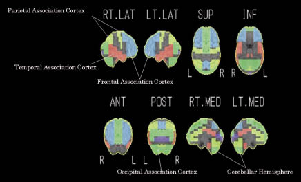
We used the stereotactic VOI template included in the 3D-SSP programme to compare the averaged 123I-IMZ activity between MTBI and controls in the frontal association cortex, temporal association cortex, parietal association cortex, occipital association cortex, and cerebellar hemisphere. A predefined set of VOIs covering these areas is illustrated in Fig. 1.

Fig. 1. Stereotactic volume of interest (VOI) template for the averaged area map on 3D-SSP analysis. Software iNEUROSTAT++ ++ (Nihon Medi-Physics Co., Ltd, Tokyo, Japan) was used for VOI analysis.
In addition, right and left differences in 123I-IMZ uptake in the areas previously described were compared among the 9 subjects with MTBI. To exclude the effect of crossed cerebellar diaschisis, right and left differences when the pixel value was normalized by the average count of the cerebellum on the same side were compared. We used computer software iNEUROSTAT++ for VOI analysis.
Statistical analysis
The results are given as means with SD. Differences in the left-to-right ratios of receptor uptake between patients and controls were tested using the unpaired t-test. Hemispheric differences in benzodiazepine receptor uptake were tested using the paired t-test. A probability threshold of p < 0.05 was assumed to represent a significant change.
RESULTS
Profiles of subjects at the time of examination
Values of the FIMTM at the time of the examination and results of the neuropsychological tests are shown in Table I. All patients had abnormal results in at least one of the subsets of these evaluation methods. Mean FIM motor score was 91.0 (SD 0), mean FIM cognition score was 32.4 (SD 1.0) (range 30–33), mean verbal intelligence quotient of WAIS-R was 100.9 (SD 17.6) (range 79–121), mean performance intelligence quotient of WAIS-R was 105.9 (SD 23.3) (range 68–136), and mean full intelligence quotient of WAIS-R was 105.9 (SD 19.4) (range 84–128).
|
Table I. Profiles of patients with mild traumatic brain injury |
||||
|
At time of injury |
Time since injury, years |
At time of examination |
||
|
Age, years/ gender |
Cause of injury |
Coma level |
Self-reported symptoms |
|
|
22/M |
Traffic accident, bicycle riding |
GCS/15 |
2 |
Headache, dizziness, fatigue, memory problems |
|
31/M |
Traffic accident |
GCS/14 |
4 |
Headache, dizziness, fatigue, memory problems |
|
52/M |
Traffic accident, as a passenger |
GCS/15 |
12 |
Dizziness, fatigue, poor concentration, memory problems |
|
51/F |
Traffic accident, as a passenger |
GCS/15 |
7 |
Headache, dizziness, fatigue, anxiety |
|
49/F |
Traffic accident, as a passenger |
GCS/15 |
10 |
Headache, dizziness, fatigue, anxiety |
|
31/M |
Traffic accident, motorcycle riding |
GCS/15 |
3 |
Headache, dizziness, fatigue, memory problems |
|
33/F |
Traffic accident, motorcycle riding |
GCS/13 |
9 |
Dizziness, fatigue, poor concentration, memory problems |
|
57/F |
Traffic accident, bicycle riding |
GCS/15 |
3 |
Dizziness, fatigue, poor concentration, memory problems |
|
38/M |
Traffic accident |
JCS/2 |
7 |
Headache, dizziness, fatigue, memory problems |
|
M: male; F: female; GCS: Glasgow Coma Scale; JCS: Japan Coma Scale. |
||||
SPECT analysis
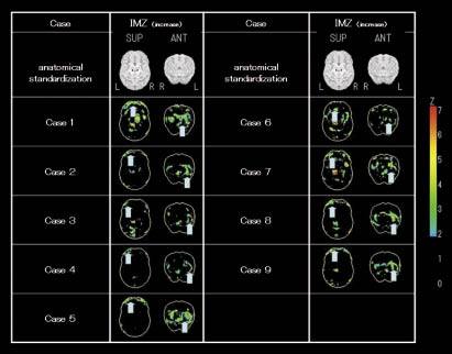
The degree of increase in blood flow is shown as Z-values ((mean in the normal group – patient’s value)/SD in the normal group). The results of the analysis are shown as a projection chart on a 3D brain, thus the position of increased blood flow can easily be located. Because of this advantage, this method is useful in daily clinical diagnosis. In this study, the region of the brain with a Z-value corresponding to +2.0 was considered to be a site where benzodiazepine receptor uptake increased significantly (the region where benzodiazepine receptor uptake was more than 2.0 SD above the control data was indicated as a site with abnormal benzodiazepine receptor uptake). We found that in all of the patients with MTBI, benzodiazepine receptor uptake in the prefrontal cortex was significantly higher (more than 2 SDs) than in the controls (Fig. 2).

Fig. 2. Regional increases in benzodiazepine receptor uptake demonstrated by 123I-iomazenil single photon emission computerized tomography (SPECT). The brain region with a Z-value corresponding to +2.0 was considered to be a site where benzodiazepine receptor uptake increased significantly. The region where benzodiazepine receptor uptake was more than 2.0 standard deviations above the control data was indicated as a site with abnormal benzodiazepine receptor uptake.
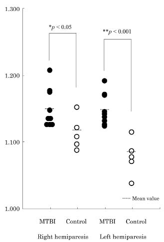
We also found significantly higher frontal association cortex to averaged global brain activity ratios in the MTBI patients than in the controls in both hemispheres (Fig. 3).

Fig. 3. Scatter plots of the frontal association cortex-to-averaged global brain activity ratios in patients with mild traumatic brain injury (MTBI) and in control subjects.
In the patients with MTBI, there was a greater uptake ratio of 123I-iomazenil in the right frontal association cortex than in the left (n = 9, p < 0.05) (Table II). On the other hand, we did not find any significant laterality of uptake ratio in any other brain area except the cerebellar hemisphere (Table II).
|
Table II. Mean (standard deviation (SD)) regional 123I-iomazenil uptake in patients with mild traumatic brain injury (MTBI) (n = 9). In the patients with MTBI, uptake ratio of 123I-iomazenil in the right frontal association cortex was greater than in the left. Figures are normalized to the mean global brain count density |
|||
|
Region of interest |
Right |
Left |
p |
|
Frontal association cortex |
1.150 (0.029) |
1.148 (0.023) |
0.6609 |
|
Frontal-to-cerebellar ratio |
1.352 (0.124) |
1.314 (0.106) |
0.0416* |
|
Temporal association cortex |
1.138 (0.032) |
1.136 (0.045) |
0.8092 |
|
Temporal-to-cerebellar ratio |
1.390 (0.160) |
1.331 (0.145) |
0.1503 |
|
Parietal association cortex |
1.180 (0.068) |
1.161 (0.059) |
0.2031 |
|
Parietal-to-cerebellar ratio |
1.336 (0.080) |
1.298 (0.079) |
0.0577 |
|
Occipital association cortex |
1.201 (0.060) |
1.210 (0.041) |
0.4876 |
|
Occipital-to-cerebellar ratio |
1.409 (0.102) |
1.348 (0.097) |
0.2471 |
|
Cerebellar hemisphere |
0.078 (0.062) |
0.855 (0.065) |
0.0336* |
|
*significant. |
|||
DISCUSSION
As sequelae following traumatic brain injuries, cognitive dysfunction, decreases in memory, attention, and concentration, changes in personality and emotional or behavioural disorders are well known. The most common areas of brain contusion in traumatic brain injuries are the frontal and temporal lobes. However, cognitive dysfunction similarly exists in patients with MTBI in whom the absence of regional lesions is clearly shown on diagnostic imaging of the brain. Although accounting for more than 70% of cases of head injury, MTBI has lacked attention by neurosurgeons and neurologists alike (6). Although positron emission tomography provides information on cerebral metabolism and has shown promise in MTBI (9), it is not widely available, not regularly reimbursed, and is more expensive than SPECT. The discovery of significant changes in relative cerebral blood flow in patients with MTBI by various researchers (10–12) makes SPECT a promising tool for the evaluation of MTBI. Jacob et al. (13) evaluated 136 patients with MTBI who underwent initial SPECT within 4 weeks after trauma and were followed at 3, 6, and 12 months after injury. They concluded that normal SPECT findings are a reliable tool to exclude clinical sequelae of mild injury. Also, at 12 months post-injury, a positive SPECT study is also a reliable predictor for clinical outcome (14). It has been suggested that the earlier the SPECT is performed after MTBI, the greater the number of lesions will be detected (15). However, in many cases, sequelae of MTBI are diagnosed a long time after the injury.
In this study, we detected abnormal benzodiazepine receptor uptake in the prefrontal cortex in all cases with MTBI by 123I-iomazenil SPECT. Kuikka et al. (16) demonstrated focally increased benzodiazepine receptor uptake in the right prefrontal cortex in patients with panic disorder, which supported the idea that the difficulty in extinguishing a learned phobic disorder is associated with deterioration of information processing in the prefrontal cortex. Clinically, the differential diagnosis between MTBI and panic disorder is sometimes difficult or it may be that these diseases overlap each other. The present study provides evidence that 123I-iomazenil SPECT is a useful tool for diagnosing both disorders. It is thought that a significant impact, such as a traffic accident, accompanied by rotary acceleration destroys the function of the prefrontal area of cerebral hemisphere first. Hashimoto et al. (17) reported a case of a man who had MTBI as a result of an accident and that some fibres from the corpus callosum towards the frontal cortex were noted on tractography of magnetic resonance diffusion tensor imaging. Therefore, the laterality of benzodiazepine receptor uptake in the prefrontal cortex in patients with panic disorder and MTBI may be associated with weakness of neural function in response to the impact, especially in the prefrontal cortex (17). The results of the past study and this SPECT study may explain the symptoms after MTBI by destruction of neural function in this area. On the other hand, some limitations of this study must be mentioned. One is the too small sample size, another is the long time-span between injury and examinations, ranging from 2 to 12 years, and the other is the possible confounding of both neuropsychological symptoms and SPECT data. And we must also consider the possible impact of confounds of both the neuropsychological and the SPECT data by factors such as depression, anxiety or pain.
To summarize, the present study provides evidence that some neural function is altered in the prefrontal cortex in patients with MTBI. Our SPECT study demonstrated focally altered benzodiazepine receptor uptake, particularly in the prefrontal cortices in patients with MTBI.
ACKNOWLEDGEMENT
The authors thank Dr M. Uchiyama and Dr S. Ogi for specialist advice on SPECT.
REFERENCES
