OBJECTIVE: To investigate the co-ordination between reaching, ground reaction forces and muscle activity in standing children with severe spastic diplegia wearing dynamic ankle-foot orthoses compared with typically developing children.
DESIGN: Clinical experimental study.
SUBJECTS: Six children with spastic diplegia (Gross Motor Function Classification System level III-IV) and 6 controls.
METHODS: Ground reaction forces (AMTI force plates), ankle muscle activity (electromyography and displacement of the hand (ELITE systems) were investigated while reaching for an object.
RESULTS: For the children with severe spastic diplegia who were wearing dynamic ankle-foot orthoses, co-ordination between upward and forward reach velocity differed regarding the temporal sequencing and amplitude of velocity peaks. During reaching, these children lacked interplay of pushing force beneath the reach leg and braking force beneath the non-reach leg and co-ordinated ankle muscle activity, compared with controls.
CONCLUSION: The results suggest differences in reach performance and postural adjustments for balance control during a reaching movement in standing between children with spastic diplegia Gross Motor Function Classification System level III–IV, wearing dynamic ankle-foot orthoses compared with typically developing children.
Key words: spastic diplegia, orthoses, reaching, kinetics, kine matics and postural adjustments.
J Rehabil Med 2007; 39: 715–723
Correspondence address: Annika Näslund, Department of Health Science, Luleå University of Technology, SE-971 87 Luleå, Sweden. E-mail: annika.naslund@ltu.se
Submitted January 18, 2007; accepted June 27, 2007.
INTRODUCTION
Reaching and grasping are basic and important upper extremity multi-joint movements for activities of daily living. According to the systems theory of motor control, specific neural and musculoskeletal subsystems contribute to the co-ordination of multi-joint movements (1).
Children with normal motor development develop inter-joint co-ordination during the first years of reaching (2). The development of reach and grasp emerges progressively throughout early infancy and childhood, whereas the abilities to locate the object in space and transport the arm are thought to be innate and are present in a rudimentary form at birth (3). Normal reaching movement engaging more than one joint is characterized by smooth, approximately bell-shaped velocity profiles and straight trajectories (4), indicating that reaching mostly is pre-programmed in advance (5). The velocity of hand transport varies with target distance and the grip is pre-shaped according to the shape and size of the object (6).
Children with cerebral palsy (CP) experience movement disorders (motor impairment) due to velocity-dependent increases in tonic stretch reflexes (spasticity), muscle weakness, excessive co-activation of antagonist muscles and increased stiffness around joints (7). Postural dysfunction is a contributing factor to problems with functional skills such as reaching movements (8, 9). The reaching movement is characterized by multi-joint dys-co-ordination leading to abnormal movement trajectories (1).
Children with spastic CP often use ankle-foot orthoses to improve standing and walking as well as to facilitate function (10) and for obtaining optimal postural control (11). In Norrbotten, Sweden (a province in northern Sweden), dynamic ankle-foot orthoses (DAFOs) have been used as an adjunct to physiotherapy for children with spastic diplegia in order to improve sitting, standing, walking as well as to improve arm and hand function. Research has suggested positive effects in children with spastic diplegia who wear DAFOs, such as a more even weight distribution (12) and the use of anticipatory postural adjustments (13) in standing.
Few studies have compared kinematic characteristics of reaching movements in children with CP with typically age-matched developing children (14, 15), and to our knowledge no study has investigated the reaching movement in children with severe spastic diplegia in standing with some support. Because reaching is usually a forward and upward displacement of the hand, there is a lack of knowledge about the temporo-spatial interaction between both movement dimensions. The aim of this study was to investigate the temporal and spatial parameters of reaching movements and the co-ordination among hand movement, ground reaction forces and muscle activity in standing in children with severe spastic diplegia (as classified according to Gross Motor Function Classification System (GMFCS) I–V, at level III–IV (16)) who were wearing DAFOs. A further aim was to compare the findings with unimpaired controls.
METHODS
Subjects
All children (aged 5–12 years) with spastic diplegia who, at the time of the study (2001), were using DAFOs (n = 17) in the County of Norrbotten were invited to participate. Patients’ charts and a special register of patients with CP contained the information needed to verify diagnosis, age and orthosis.
Informed consent was obtained from the parents for the research project and the assurance of confidentiality and their right to refuse or withdraw was clearly stated. Eight parents gave their informed consent for their child to participate in the study. Classification of gross motor function was performed according to GMFCS I–V (16). According to GMFCS criteria one participant was classified at level II and 7 at level III–IV. One of these 7 participants had to be excluded after the test procedure due to pain, since he had grown out of the DAFOs. The 6 participants at level III–IV thus formed the study group (CP). The children with spastic diplegia had severely restricted mobility and were dependent on walking aids and/or living aids (level III–IV). At the time of the study, the children were between 5 and 12 years of age (mean age 7.8 years) and had been using DAFOs for an average of 2 years. Individual data are presented in Table I. Eight children within the same age group and with no known physical problems served as a control group. The study was approved by the ethics committee at Umeå University, Sweden (Dnr-00-022).
| Table I. Main clinical features in children with cerebral palsy with spastic diplegia treated with dynamic ankle-foot orthoses (DAFOs) |
| Subject | Sex | Age (years) | Weakest side | Surgery | Type of surgery | DAFO start | GMFCS |
| 1 | F | 5 | right | 1998 | B,C,D | 1998 | III |
| 2 | M | 9 | left | 1998 | A,C | 1997 | IV |
| 3 | M | 7 | right | 1998 | A,B,C | 1997 | III |
| 4 | M | 6 | right | none | | 1997 | IV |
| 5 | M | 12 | right | 1998 | B,C,E | 1997 | III |
| 6 | F | 8 | left | 1996 | B,C,D | 1997 | III |
| A: iliopsoas lengthening; B: distal medial hamstrings lengthening; C: adductor lengthening; D: Achilles tendon lengthening; E: peroneus lengthening. GMFCS: Gross Motor Function Classification System I–V. |
Instruments and procedure
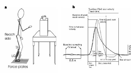
The participants stood in front of a table adjusted to waist height, with each foot on a separate force plate (AMTI, Advanced Mechanical Technology Incorporated, model MC818-6-1 000; size 457 × 203 mm; accuracy 0.25 N) indented into the floor. The force plates registered the forces along the 3 orthogonal axes. The participants required a feeling of security during the task performance, which was provided either by placing their left hand on the edge of the table or by a minimal assistance provided by their parent’s hand on the pelvis. The participants were thus not lifted or supported during the task. Subject 1 was given assistance at the pelvis, subjects 2, 3 and 4 alternated between hand and pelvis assistance, and subjects 5 and 6 used hand assistance. In the present study the analysis is limited to the anterior-posterior ground reaction forces, which we expected to change in association with the reaching task. A three-dimensional optoelectronic movement analysis system (ELITE, Elaboratore di immagini televisive BTS, Milan, Italy) (17) with 2 charge-coupled device cameras was placed on the participant’s right side at a distance of 2 m. This system was used to register the position and displacement of the hand from a hemisphere-shaped reflective wrist marker (diameter 15 mm) that was fastened with adhesive tape to the right wrist. The calibrated volume was 2 cubic m, with an accuracy for that volume of 0.78 mm3. Bilateral muscle activity of the tibialis anterior (TA) and lateral gastrocnemius (LG) were recorded by surface electromyography (EMG, Bagnoli-8, Delsys, Boston, MA). The electrodes were a differential type pre-amplified with a gain of 10, and an inter-electrode distance of 10 mm (DE-02, size 23 × 17 mm). The surface electrodes were attached with adhesive tape over the respective muscle bellies. Kinematic, EMG and force plate signals were recorded simultaneously.
The task was to use the right hand to reach, at self-selected velocity, for a cup filled with sweets and place it on the table. The cup was placed at eye level. The experimental set-up is shown in Fig. 1(a). The CP group and controls performed the task with the cup placed at a distance of an arms’ length plus 10%. The CP group performed the task while wearing DAFOs and shoes fitted for DAFOs, since this was the shoe support they commonly used for all activities during the day. The controls performed the same task wearing regular shoes. To encourage the children to complete the task, they were told to take a sweet for each trial that was completed and to save it for later.
Data collection analysis
Kinematic, force plate and electromyographic signals were simultaneously recorded during 7 sec, starting one sec before each trial, in order to obtain baseline values on the ELITE computer and the SC/ZOOM computer. The sampling frequencies were 100 Hz for the wrist marker and the force plate signals and 800 Hz for the EMG. The signals were digitized with an A/D converter at 12-bit resolution and stored on SC/ZOOM (a flexible laboratory computer system; Department of Physiology, Umeå University) and on the ELITE computer for further analysis. The raw EMG signals were amplified by a gain of 200 and band-pass filtered between 10 Hz and 1 kHz. EMG signals were processed using the root mean square with 1.25 millisec sampling interval for rectification in order to differentiate signal artefacts. The force plate data were collected, together with the EMG signals on SC/ZOOM and simultaneously with the kinematic data on the ELITE computer. The force plate and kinematic data were transformed into ASCII files. By using Axograph (Axon Instruments, Inc., Union City, CA, USA), a Macintosh-based software package that allows custom-made-semi-automatic routines, the wrist velocity was used for defining time events and peak amplitudes of the forward and upward reach movement. The measured kinematic variables are indicated on a representative example of the reach velocity profile of the upward movement (Fig. 1(b)). The temporal course of the reaching movement was determined by the segmentation of the velocity profiles. All temporal events were defined with respect to the onset of reach movement, which was identified from cursor read-outs of a continuous increase in upward and forward velocity of the wrist marker. That instant was set to zero time and referred to as onset of reach. The onset of upward and forward velocity occurred simultaneously, and the upward movement ended prior to the forward movement. Therefore, in this study, end of reaching time was defined at the first instant in time at which the forward wrist marker velocity declined to zero. In the literature, movement time for reaching movements is usually defined as the time between onset of hand movement and onset of object displacement, i.e. task completion (4). However, in the CP group some subjects had difficulties with grasping and lifting the object, resulting in a large variation of time between end of the hand transport component and the grasping lifting component. In this study, the reach time is defined as between onset and stop of the velocity trace of the forward movement. The time between onset and peak velocity identified the acceleration phase and the time between peak velocity and zero velocity identified the deceleration or braking phase of the reach velocity profiles.
Fig. 1. (a) Experimental setup for the reach task showing the force plates and muscles of electromyography recording of the reach and non-reach side. (b) Time traces of the upward and forward velocity of 1 reach trial illustrating the different parameters analysed. Acc.: acceleration; Dec.: deceleration; fwd:, forward; upwd: upward; TA: tibialis anterior; LG: lateral gastrocnemius.
The time elapsed between onset of velocity of wrist marker forward and upward movement to the instant of zero velocity of the wrist forward movement was standardized as 100% reach time. This 100% reach time, as well as the upward movement, were divided into 2 phases; acceleration and deceleration. Five reaching trials were analysed for each subject, resulting in the analysis of 30 trials for each group.
The mean amplitudes of the anterior-posterior forces were calculated over the time window of the identified acceleration and of the deceleration phase of the forward displacement of the wrist marker.
EMG responses for each muscle are reported as a ratio of the background level of activity, i.e. the mean amplitude of baseline EMG calculated over a 500 millisec window before onset of reach velocities. The mean amplitude of EMG response of each muscle was measured over the time window of the acceleration and deceleration phases and expressed in relative units to the calculated baseline amplitude.
Statistical analysis
All statistical analyses were performed using STATISTICA for Windows (StatSoft Inc., Tulsa). The means of 5 reach trials constituted the result for each child. The kinematic data are reported as mean (standard deviation (SD)) of all individual means of the group. However, for both force plate and EMG analyses, the group median and range of the individual means is reported because the group mean differed significantly from the median value. Significance level was set at p < 0.05. Non-parametric statistics were used to make comparisons within groups using Wilcoxon’s matched-pairs test. The Mann-Whitney U test was used to identify differences between the groups.
RESULTS
Reach performance
To be able to reach the cup, a displacement of the hand in the upward and forward direction was required. Both groups were able to complete the task. However, the quality of the reach differed in several aspects.
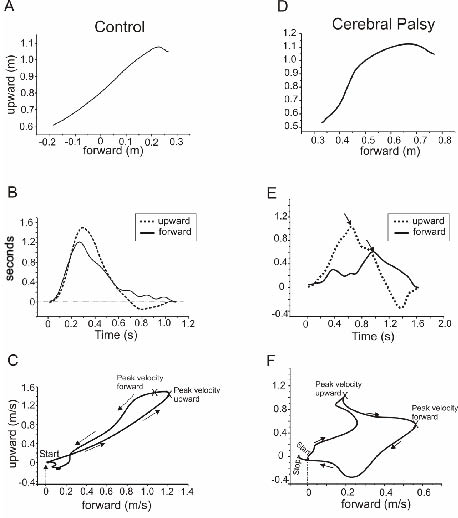
In Fig. 2, the trajectory of the wrist marker, the temporal sequencing and the relation of the amplitude scaling between forward and upward wrist velocity are illustrated for one control and one child with CP.
The trajectory displaced by the control child is smooth and approximately straight, confirming a distinct co-ordination between upward and forward wrist movement (Fig. 2A). The specific temporal sequencing between upward and forward velocity shows a steep slope of both until peak velocity (Fig. 2B). The velocity behaviour regarding the interaction of the scaling amplitude is visualized by plotting the velocity of the forward movement against the velocity of the upward movement (Fig. 2C). The resulting phase plane graph typifies the path of a lasso throw.
The wrist trajectory of the subject with CP suggests a more segmented interaction between forward and upward displacement (Fig. 2D), which is confirmed in the temporal sequencing of the velocity peaks (Fig. 2E) and in the scaling of the forward and upward velocity (Fig. 2F).
Fig. 2. X–Y plot of a single trial of the reach trajectory of the wrist marker of (A) one control and (D) one child with cerebral palsy with spastic diplegia. For the same trials, the velocity profiles of the upward and forward movement are shown (B, E). Note the difference in time of the velocities peak amplitude. The X–Y plot of the reach forward and upward velocity (C, F) illustrating the co-ordination between the forward and upward velocity path. Arrows indicate path direction and × indicates instant of the peak forward and upward velocity.
In the following, we analysed in detail the velocity profiles of wrist forward and upward movement for identifying basic motor control principles of planning and performing this task-specific reach in children with more severe CP.
Temporal course of the reach movement
Reach movement time. The duration of the wrist forward and upward movement was significantly longer for the CP participants (1626 (SD 296) millisec, 1181 (SD 247) millisec, respectively) compared with the controls (1175 (SD 158) millisec, 766 (SD 126) millisec, respectively) (p = 0.025, p = 0.006). The upward movement time compared with forward movement time was significantly shorter in both groups (CP group, p = 0.043; controls, p = 0.028).
Time of peak velocity. Peak velocity of forward and upward wrist movement was reached close to each other in the controls, while in the CP group there was a larger delay between velocity peaks. In the control group, forward peak velocity was reached 49 (SD 31) millisec prior to upward in 23 out of 30 trials. In the remaining 7 trials peak time of the forward velocity was delayed compared with upward (185 (SD 58) millisec) in 3 subjects (2 subjects in 3 trials and one subject in one trial). In the CP group the peak time of forward velocity occurred 182 (SD 127) millisec prior to the peak time of upward velocity in 10 out of 30 trials. In the remaining 20 trials peak time of upward velocity preceded the peak time of forward velocity by 347 (SD 105) millisec.
Duration of acceleration and deceleration phase. For the CP group the acceleration phase of the forward (719 (SD 213) millisec) and upward (584 (SD 91) millisec) movement phases as well as the deceleration phase of the upward movement (596 (SD 170) millisec) were significantly longer (p = 0.016, p = 0.01, p = 0.006) compared with controls (366 (SD 104) millisec, 360 (SD 60) millisec, 406 (SD 80) millisec). However, the deceleration time of the forward movement was about the same for both groups (CP group; 906 (SD 324) millisec, controls; 809 (SD 152) millisec, p = 0.426).
Acceleration and deceleration phases in percentage of reach time. The acceleration phase of the upward movement was significantly longer in the CP group (37% (SD 5%)) compared with controls (31% (SD 3%)) (p = 0.024). However, this was not the case for the deceleration phase (CP group; 37% (SD 10%), controls; 35% (SD 6%), p = 0.748). In the forward movement the duration of the acceleration phase (CP group; 46% (SD 15%), controls; 31% (SD 8%)), and the deceleration phase (CP group; 54% (SD 15%), controls; 69% (SD 7%)) were not significantly different (p = 0.065, p = 0.065, respectively) between the groups.
In the controls there was a significant difference in duration of the forward acceleration compared with the deceleration phase (p = 0.028), however this was not the case in the CP group (p = 0.753). There was no difference in duration of the upward acceleration phase compared with the deceleration phase in the 2 groups (CP; p = 0.0753, control; p = 0.116). The upward movement ended in the CP group after 73% (SD 14%) and in the control group after 65% (SD 8%) of reach time, (p = 0.376).
Amplitude of reach velocity. Peak velocity of forward and upward movement was significantly lower in the CP group (0.469 (SD 0.213) m/sec, 0.900 (SD 0.425) m/sec, respectively) compared with controls (1.144 (SD 0.265) m/sec, 1.499 (SD 0.32) m/sec, respectively) (p = 0.004, p = 0.025). For both groups, the peak velocity of the upward movement was significantly higher compared with the forward movement (CP, p = 0.028; controls, p = 0.028).
Postural adjustments
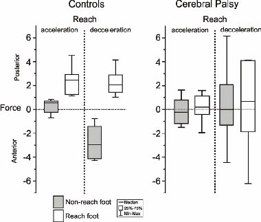
Interplay of the anterior/posterior ground reaction forces between reach and non-reach sides. Reaching was associated with changes in the anterior/posterior ground reaction forces beneath the reach and non-reach foot (Fig. 3). In the controls there was a distinct interaction between the anterior/posterior forces between the feet regarding the acceleration and the deceleration phase of the reach. During the acceleration phase the anterior/posterior force beneath the foot on the non-reach side oscillated around zero, while the reach side foot applied a posterior directed force (pushing) of about 2 N (2.42). During the deceleration phase the reach foot maintained a similar amount of posterior directed force as during the acceleration phase, while the non-reach side foot applied an anterior directed force of about 3 N (2.94), indicating a braking force. This suggests in normal subjects interplay between the reach side during the acceleration (pushing) and non-reach side during the deceleration (braking force) in order to reach forward towards the object.
The CP group lacked interplay between the anterior/ posterior forces during the acceleration and deceleration phases as both feet showed an oscillation around zero. The median force amplitude was about the same, around zero, during both phases. The range of the anterior/posterior forces was much larger during the deceleration phase for the CP group compared with the controls, indicating difficulties in stopping the forward reaching movement (Fig. 3).
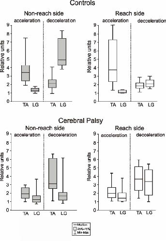
Fig. 3. Box and whisker plots of group median of individual mean amplitudes of anterior/posterior force during the reach acceleration and deceleration phase of the reach and non-reach foot plotted for the control and the children with cerebral palsy with spastic diplegia.
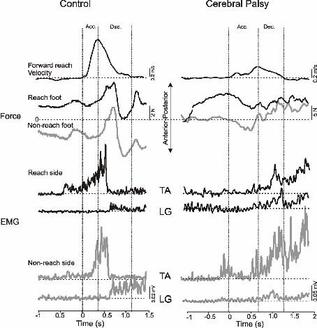
Co-ordination of muscle activity between reach and non-reach sides. The co-ordination of ankle muscle activity in TA and LG with respect to the reaching phases and the anterior/ posterior forces is illustrated in Fig. 4 for one child with CP and one control subject.
Fig. 4. Time traces of a single trial of forward reach velocity, of anterior/posterior forces and ankle muscle electromyography (EMG) on the reach (black traces) and non-reach side (grey traces) for one child of the control and the children with cerebral palsy with spastic diplegia. Time zero is indicated at onset of reach. The dashed vertical lines showing onset, time of peak and stop of forward velocity, define the time frame of the acceleration (acc.) and deceleration (dec.) phase. TA: tibialis anterior; LG: lateral gastrocnemius.
During the acceleration phase of the forward movement compared with baseline, the controls increased TA activity on the reach side (3.77) and on the non-reach side (3.45). The LG activity remained almost unchanged (reach side 1.11; non-reach side 1.32). During the deceleration phase compared with the acceleration phase the controls increased significantly the activity in the LG on both sides (reach side, 2.11, p = 0.028; non-reach side, 4.92, p = 0.028), while the TA activity (reach side, 1.88, p = 0.075, non-reach side, 2.16, p = 0.116) decreased. Note, the LG activity is much larger on the non-reach side compared with the reach side. During the acceleration phase the CP group increased TA (1.96) and LG (1.4) activity similarly on the reach side and on the non- reach side TA (1.88) and LG (1.3). During the deceleration phase compared with the acceleration phase both the TA and LG activity increased significantly on both sides (TA; reach side 3.63, p = 0.028, non-reach side 3.15, p = 0.028; LG; reach side 3.43, p = 0.046, non-reach side 1.68, p = 0.046) (Fig. 5).
Fig. 5. Box and whisker plots of group median of individual mean amplitudes of ankle muscle electromyography (expressed in relative units, see methods) during the reach acceleration and deceleration phase of the reach and non-reach side, shown for the control and the children with cerebral palsy with spastic diplegia. TA: tibialis anterior; LG: lateral gastrocnemius.
DISCUSSION
The present study examined reaching quality with respect to the interaction of forward and upward transport of the hand in space and associated postural adjustments during supported standing in children with severe spastic diplegia compared with independently standing controls. The results suggest that the co-ordination between upward and forward reach velocity differed regarding the temporal sequencing (phasing) and amplitudes of velocity peaks for the children with CP compared with the controls. The postural adjustments during the reach in controls were characterized by distinct increased TA activity on the reach side during the acceleration phase of the forward velocity and of increased LG activity on the non-reach side during the deceleration phase. Despite the child’s support of the non-reaching hand and/or the parent’s pelvis support for standing during reach performance the CP group showed co-contraction of TA and LG during the acceleration and deceleration phase on the reach and non-reach sides.
Reach kinematics during supported standing
Graphic representations of kinematic measures, in our case the velocity profiles of forward and upward movement, yield an immediate “snapshot” of the quality of the reach. This was demonstrated in the present study by the use of velocity time traces and velocity-velocity plots (Fig. 2). It may be argued that isolated analysis of the hand marker is of limited value from a functional point of view. It must be considered, however, that optimal control of upper limb movement is critically dependent on the temporal and spatial co-ordination of many joints and is reflected in the kinematics of the end point, i.e. hand marker (18). With single joint analysis (elbow) and graphic representations of such phase plane and angle-angle plots, Ramos et al. (19) nicely demonstrated the dys-co-ordination of guided reaching in children with Friedreich’s ataxia.
While generally, for controls in the majority of trials, the time of peak forward velocity was some 10 millisec earlier than the time of peak upward velocity, in the CP group upward velocity usually peaked prior to forward velocity. One possibility is that children with CP plan upward and forward displacement in sequence, i.e. in a stepwise manner, resulting in a reach trajectory with a steep initial and thereafter a more flattened slope. Also, lifting the hand upward by flexing the elbow as well as internal rotation of the shoulder and forearm pronation (20) is a clinically common pattern seen in children with CP, which could be less challenging for postural control than lifting the hand with straight elbow. An important feature of reach performance is that the end-point (wrist marker in our study) typically follows bell-shaped or right-skewed bell-shaped velocity profiles (see (21) for review). Bell-shaped velocity profiles reflect that start and stop of the reach are planned prior to movement onset. For structuring early infant reaching, von Hofsten (22) described reaching in terms of movement units, which had been described by Jeannerod (6). The acceleration trace of a bell-shaped velocity profile is single peaked and reflects 1 movement unit consisting of one positive curve (acceleration phase), switching to zero at the instant of peak velocity, and of one negative curve (deceleration phase). Several movement units suggest corrections during the reach path. Infants starting to reach consistently have about 3–7 movement units, while children of about 12 years of age, like adults, have one movement unit (4). Interestingly, the velocity profiles of the upward movement, but not of the forward movement, were single peaked in the controls aged 5–12 years (see Fig. 2). Additional peaks were seen during the deceleration phase of the forward movement, suggesting that in this task the controls had to make corrections towards the end of the reaching path. The CP group had less correction (peaks) during the acceleration phase of the upward movement, while forward velocity profiles in general were multi-peaked. This finding suggests that the acceleration phase of the upward movement was in agreement with the planned trajectory, but that the deceleration of the reach in order to grasp the object needed correction. In agreement with other studies on reaching in sitting, the CP group showed significantly longer reach movement duration than the controls (14, 15, 19). In the reach investigated in our study the duration of the acceleration phase of both upward and forward velocity was about the same (30%) in controls while the duration of the deceleration phase of the forward velocity was significantly longer compared with the upward velocity. This suggests that the profile of the upward velocity was bell-shaped, while the profile of the forward velocity was bell-shaped skewed to the right. Bell-shaped velocity profiles suggest similar duration of acceleration and deceleration phase. In the CP group the durations of the acceleration and deceleration phases in percent of upward and forward velocity time, respectively, were almost similar. We may assume that the controls during the deceleration of the forward reach shaped the hand for grasping the object, because immediately after the end of the forward velocity the object was lifted. As reported in the methods we defined reach time according to when forward velocity was zero and not when the object was grasped and started to move. The CP group needed several seconds after the end of the forward velocity until the object was lifted. It seems, therefore, that the children with CP performed the hand transport and hand shaping for grasping in a serial manner, rather than in parallel as in the controls. However, further studies are needed to confirm this.
Influence of pelvis or hand support on postural adjustments in association with reach performance
All children in the CP group were wearing specially designed dynamic ankle-foot orthoses DAFO supporting the foot arches, the subtalar joints and the forefoot, while allowing small movements in the mid- and fore-foot, thus making somatosensory feedback possible for balance control (11). In our study, standing support provided by the parent on the pelvis and/or by contact of the non-reach hand on the table was necessary during reach performance. Task-specific reaching and grasping occurs with respect to the reference frame of body posture (23). Supported standing as seen in this study may suggest that the child with CP could rely on the body posture for planning the reaching trajectory. While all children with CP were able to perform the task, the quality of reaching differed in some aspects compared with controls. Three of the children with different support conditions were part of a previous study showing onset of centre of pressure (CoP) displacement prior to onset of reach performance (13). This result suggests that because reaching is a forward-oriented movement, assistive standing with pelvis support by the parent or hand support of the child did allow anticipatory postural behaviour. These findings were opposite to the well-known fact based on Cordo & Nashners’ (24) research of healthy adults that when lifting the arm with concomitant contra-lateral hand support, the anticipatory leg muscle activity was suppressed. Studies on postural control of adults with light touch contact of the fingertip have demonstrated that somatosensory cues are a powerful orientation reference for improved control of upright stance (25). In the present study, however, we focused on the postural adjustments during the reach and not on improved control of upright stance.
Controls showed a pronounced reciprocal increase in TA activation during the acceleration phase of the reach both on the reach and non-reach sides (Figs 4 and 5). The CP group also had increased TA activity, although the proportion of increase compared with the antagonist LG activity was much smaller compared with the controls, suggesting a less reciprocal agonist/antagonist activation pattern. Activation of TA during the reach acceleration phase suggests that the body behaves like an inverted pendulum, where the trunk is following the forward arm movement. These findings are in agreement with Tyler & Karst (26) suggesting those in standing position the ventral muscles, such as TA, assist the reaching movement in adults instead of resisting the perturbation caused by the reach. In adults reaching while standing, anticipatory TA activity is known to induce a posterior displacement of CoP for creating the angular momentum for moving canter of mass (CoM) forwards (27) when the target is placed beyond arm length. In agreement with Kaminski & Sampkins (28), we showed that TA activity continued during the acceleration phase of the reach with a reaching distance of arm length plus 10%. In controls, the TA activity was in agreement with the applied posterior force beneath the reach side foot, resulting in a propulsive impulse forward (Fig. 4), while the CP group maintained the anterior/posterior forces around zero beneath both feet.
During the deceleration phase of the reach, controls activated LG on the non-reach side and applied a forward-directed force beneath the foot, resulting in a force braking movement. Thus, controls showed distinct inter-limb co-ordination of postural reach specific behaviour between the reach and non-reach side, while the CP group lacked this kind of adjustment/co-ordination between sides. In order to stiffen the joint for reducing the degrees of freedom (28), the CP group functioning at GMFCS level III–IV demonstrated ankle joint co-activation of agonist and antagonist muscles illustrating an immature muscle activation pattern according to Konczak et al. (2). This is known to occur in the early stages of postural development when forces linked to a specific motor task have not been integrated in motor performance (29). Our results might imply that the lack of inter-limb co-ordination of muscle activity and ground reaction force pattern can be regarded as a functional strategy to compensate for dysfunctional capacity in this group of children. Findings of increased recruitment of antagonist muscles in children with CP compared with typically developing children are consistent with those of numerous researchers studying postural control in standing (30, 31) and sitting (32) in response to platform perturbations. The action/reaction forces oscillations around zero during the acceleration phase, together with an increased magnitude during the deceleration phase in the CP group, could be explained by the non-selective activity in the ankle joint as well as by an increased transverse rotation of the trunk. This is in line with Ferdjallah et al. (33), who concluded through examination of CoP movement after perturbation in the anterior-posterior direction, that for postural stability, children with CP used a transverse body rotation strategy to compensate for poor ankle control. Furthermore, this compensatory strategy to keep CoM within base of support, is demonstrated in children with CP who moved their CoP more in a medial-lateral direction and less in the anterior-posterior direction compared with children with typical development while reaching forward (34). One contributing factor to disorganized muscle responses in the CP group could be the musculoskeletal alignment leading to a crouch posture. Researchers have found that a crouched posture in standing in typically developing children resulted in more co-activation of agonist-antagonist muscles during perturbation (30) and during reach from a standing position (35). A more mature inhibitory muscle pattern, a prerequisite for postural stability, is dominant from 6 years of age (36). As the results indicated, the CP group lacked in reciprocal activity. However, the degree of antagonistic activation in postural adjustments seems to be task-specific (37, 38). During postural adjustments following external perturbations while sitting or standing, children with CP used an excessive degree of antagonistic co-activation (31, 32, 37). However, during voluntary reaching in sitting, children with CP showed hardly any antagonistic co-activation in postural (neck and trunk) muscles (9, 29, 38). This is in contrast to our results that revealed that in a standing position voluntary goal-directed movement increased co-activation in the ankle muscles.
The CP group functioning at level GMFCS III-IV used DAFOs, since our intention was to provide them with reliable support base while standing. Studies suggest that DAFOs provide the child with a stable ankle joint configuration, resulting in a more stable stance posture (11) and furthermore, DAFOs demonstrate a more even weight distribution and straighter knees in this group of children (12). In clinical practice it is anecdotally assumed that ankle muscle activity is reduced using DAFOs, however, our study showed that DAFOs did not reduce the amount of ankle muscle recruitment; on the contrary, the EMG amplitude increased TA and LG activity (deceleration phase). This is in line with Burtner et al. (39) who stated that DAFOs did not decrease the muscle activity in the ankle joint during platform perturbations in children with spastic diplegia.
Our intention was, in a laboratory setting, to investigate kinetics, kinematics and muscle response in children with severe spastic diplegia. This is in contrast to other studies that have mostly focused on ambulatory spastic diplegic children. The limitations of our study are attributed to uncontrolled variables, such as degree of impairment in the upper and lower extremity, support conditions and small number of subjects. These factors might have influenced our findings regarding reach performance and postural adjustments.
In the present study the children were developmentally a heterogeneous group with a wide range of movement and posture disturbances, such as spasticity, muscle weakness, mal-alignment, asymmetry and dys-co-ordination. Therefore, we could not standardize the standing support, since the impairments varied greatly within and between GMFCS levels. However, we standardized the reaching distance to arm length + 10% according to anthropometric measures, and the object was placed at eye level. Internal validity regarding the support condition may be questioned. However, the included trials were checked for stability of the vertical forces during a sampling time of 7 seconds, which indicated that the child was standing with the same assistance throughout the whole trial. Furthermore, we did not expect support provided from behind on the pelvis to influence anticipatory and compensatory postural adjustments to a great extent, since this support did not change the base of support. The support may have influenced the forces and muscle activity, but this is doubtful as all children displayed similar force- and EMG-patterns, although with different amplitudes, during all trials.
The sample size was small and the results must be interpreted with caution and therefore might not be representative for children with spastic diplegia in general. On the other hand the small group investigated with movement and posture disorders at GMFCS level III–IV may be representative for children with spastic diplegia using DAFOs at the same GMFCS level.
The clinical implication of our study for non-ambulatory children would be that reaching should also be exercised in standing where ankle-foot muscle activity can be enhanced, which is not necessarily the case in sitting.
ACKNOWLEDGEMENTS
The authors thank the children and parents who participated in this study. Special thanks are due to Ingmarie Apel for her valuable assistance in preparing and running the experiments and analysing the data. Financial support was kindly provided by the Norrbacka-Eugenia Foundation, Stockholm, Sweden.
REFERENCES
1. Shumway-Cook A, Woollacott M. Reach, grasp and manipulation. In: Shumway-Cook A, Wollacott M, editors. Motor control, theory and practical application. Baltimore: Lippincott Williams and Wilkins; 2001, p. 447–496.
2. Konczak J, Borutta M, Dichgans J. The development of goal-directed reaching in infants learning to produce task-adequate patterns of joint torque. Exp Brain Res 1997; 113: 465–474.
3. Von Hofsten C. Eye-hand coordination in the newborn. Dev Psychol 1982; 18: 450–461.
4. Kuhtz-Buschbeck JP, Stolze H, Johnk K, Boczek-Funcke A, Illert M. Development of prehension movements in children: a kinematic study. Exp Brain Res 1998; 122: 424–432.
5. Ghez C, Gordon J, Ghilardi MF. Impairments of reaching movements in patients without proprioception. II effects of visual information on accuracy. J Neurophysiol 1995; 73: 361–370.
6. Jeannerod M. The timing of natural prehension movements. J Motor Behav 1984; 16: 199–208.
7. Crenna P, Inverno M. Objective detection of pathophysiological factors contributing to gait disturbance in supraspinal lesions. In: Fedrizzi E, Avanzini G, Crenna P, editors. Motor development in children. London: John Libbey; 1994, p. 103–118.
8. Woollacott M, Shumway-Cook A. Postural dysfunction and walking in children with cerebral palsy: What are the underlying problems and what new therapies might improve balance? Neural Plast 2005; 12: 211–218.
9. Van der Heide J, Hadders-Algra M. Postural muscle dyscoordination in children with cerebral palsy. Neural Plast 2005; 12: 197–203.
10. Knutson LM, Clark DE. Orthotic devices for ambulation in children with cerebral palsy and myelomeningocele. Phys Ther 1991; 71: 947–960.
11. Hylton NM. Postural and functional impact of dynamic AFOs and FOs in a pediatric population. J Prothetics Orthotics 1990; 2: 40–53.
12. Näslund A, Jesinkey K, Sundelin G, von Wendt L, Hirschfeld H. Effects of dynamic ankle-foot orthoses on standing in children with severe spastic diplegia. Int J Ther Rehabil 2005; 12: 200–207.
13. Jesinkey K, Näslund A, Hirschfeld H. Initiating of reaching when standing with and without DAFOs in children with spastic diplegia. Adv Physiother 2005; 7: 144–153.
14. Van der Heide JC, Fock M, Otten B, Stremmlaar E, Hadders-Algra M. Kinematic characteristics of reaching movements in preterm children with cerebral palsy. Ped Res 2005; 57: 883–889.
15. Chang JJ, Wu TI, Wu WL, Su FC. Kinematical measure for spastic reaching in children with cerebral palsy. Clin Biomech 2005; 20: 381–388.
16. Palisano RJ, Rosenbaum PL, Walter SD, Russell DJ, Wood EP, Galuppi BE. Development and reliability to classify gross motor function in children with cerebral palsy. Dev Med Child Neurol 1997; 39: 214–223.
17. Ferrigno G, Pedotti A, editors. ELITE: a digital dedicated hardware system for movement analysis via real-time TV signal processing. IEE Trans Biomed Eng BME-32 1985; 11: 943–950.
18. Latash ML. Multi-joint movement. In: Latash M, editor. Neurophysiological basis of movement. Champaign, IL: Human Kinetics; 1998, p. 179–188.
19. Ramos E, Hurvitz EA, Brown SH. Quantification of upper extremity function using kinematic analysis. Arch Phys Med Rehabil 1997; 78: 491–496.
20. Koman LA, Paterson S, Shilt JS. Cerebral palsy. Lancet 2004; 363: 1619–1631.
21. Bullock D, Grossberg S. Neural dynamics of planned arm movements: emergent invariants and speed-accuracy properties duringtrajectory formation. Psychol Rev 1988; 95: 49–90.
22. Von Hofsten C. Structuring of early reaching movements: a longitudinal study. J Mot Behav 1991; 23: 280–292.
23. Soechting JM, Flanders M. Arm movements in three-dimensional space: computation, theory and observation. Excer Sport Sci Rev 1991; 19: 389–418.
24. Cordo PJ, Nashner LM. Properties of postural adjustments associated with rapid arm movements. J Neurophysiol 1982; 47: 287–302.
25. Jeka JJ, Lackner JR. Fingertip contact influence human postural control. Exp Brain Res 1994; 100: 495–502.
26. Tyler AE, Karst GM. Timing of muscle activity during reaching while standing: systematic changes with target distance. Gait Posture 2004; 20: 126–133.
27. Kaminski TR, Sampkins S. The effects of stance configuration and target distance on reaching 1. Movement preparation. Exp Brain Res 2001; 136: 439–446.
28. Biryukova EV, Roschin VY, Frolov AA, Ioffe ME, Massion J, Dufosse M. Forearm postural control during unloading: anticipatory changes in elbow stiffness. Exp Brain Res 1999; 124: 107–117.
29. Brogren Carlberg E, Hadders-Algra M. Postural Dysfunction in children with cerebral palsy: some implications for therapeutic guidance. Neural Plasticity 2005; 12: 221–228.
30. Woollacott MH, Burtner P, Jensen J, Jasiewicz J, Roncesvalles R, Sveistrup H. Development of postural responses during standing in healthy children and children with spastic diplegia. Neurosci Biobehav Rev 1998; 22: 583–589.
31. Burtner PA, Quallis C, Woollacott MH. Muscle activation characteristics of stance balance control in children with spastic cerebral palsy. Gait Posture 1998; 8: 163–174.
32. Brogren E, Hadders-Algra M, Forssberg H. Postural control in sitting children with cerebral palsy. Neurosci Biobehav Rev 1998; 22: 591–596.
33. Ferdjallah M, Harris GF, Smith P, Wertsch JJ. Analysis of postural control synergies during quiet standing in healthy children and children with cerebral palsy. Clin Biomech 2002; 17: 203–210.
34. Liu WY, Zaino CA, Westcott SL. Anticipatory postural adjustments in children with cerebral palsy and children with typical development during functional reaching: a center of pressure (CoP) study. Ped Phys Ther 2000; 12: 218–219.
35. Thorpe D, Zaino C, Westcott S, Valvano J. Comparison of postural muscle coordination patterns during a functional reaching task in typically developing children and children with cerebral palsy. Phys Ther 1998; 78: 80–81.
36. Schmitz C, Martin N, Assaiante C. Building anticipatory postural adjustment during childhood: a kinematic and electromyographic analysis of unloading in children from 4 to 8 years of age. Exp Brain Res 2002; 142: 354–364.
37. Brogren E, Forssberg H, Hadders-Algra M. Influence of two different sitting positions on postural adjustments in children with spastic diplegia. Dev Med Child Neurol 2001; 43: 534–546.
38. Van der Heide JC, Berger C, Fock JM, Otten B, Stremmelaar E, van Eykern LA, Hadders-Algra M. Postural control during reaching in preterm children with cerebral palsy. Dev Med Child Neurol 2004; 46: 253–266.
39. Burtner PA, Woollacott M, Quallis C. Stance balance control with orthoses in a group of children with spastic cerebral palsy. Dev Med Child Neurol 1999; 41: 748–757.


