
From the 1ToNIC, Toulouse NeuroImaging Center, Université de Toulouse, Inserm, UPS, 2Ipsen Innovation, Les Ulis, 3Department of Physical Medicine and Rehabilitation and 4Department of Functional Physiological Explorations, University Hospital of Toulouse, Hôpital de Rangueil, Toulouse, France
Objective: To elucidate the adverse consequences of spasticity and spastic co-contraction of elbow flexors on motor impairment and upper limb functional limitation.
Design: A pilot case-controlled prospective observational study.
Subjects: Ten brain-injured adults, and 10 healthy controls.
Methods: The co-contraction index was computed from electromyographic recordings of elbow flexors during sub-maximal (25% Maximal Voluntary Contraction) isometric elbow extension. Spasticity was assessed with the Tardieu scale, upper limb limitation using a goniometer during active elbow extension, motor selectivity with the Fugl-Meyer Assessment for the upper limb, and motor function with the Action Research Arm Test.
Results: Greater co-contraction occurred in patients with brain injury compared with controls. In contrast to spasticity, strong associations were found between the co-contraction index, the limitation of active elbow extension, the Fugl-Meyer Assessment, and the Action Research Arm Test.
Conclusion: This pilot study suggests that spastic co-contraction rather than spasticity is an important factor in altered upper limb motricity in subjects with brain injury, leading to abnormal restricting arm movement patterns in subjects with more severe motor impairment. Practical applications directly concern the pre- and post-therapeutic evaluation of treatments aimed at improving motor skills in subjects with brain injury.
Key words: brain injury; hemiplegia; muscle hypertonia; upper extremity.
Accepted Jan 29, 2019; Epub ahead of print, Feb 15, 2019
J Rehabil Med 2019; 51: 00–00
Correspondence address: David Gasq, Toulouse NeuroImaging Center, CHU Purpan, Pavillon Baudot, place du Dr Baylac 31024 Toulouse, France. E-mail: david.gasq@inserm.fr
Spasticity and spastic co-contraction are expressions of muscle overactivity that occur in spastic paresis syndrome after a brain injury. The objective of the present pilot study was to improve our understanding of the respective adverse consequences of spasticity and spastic co-contraction on motor disability. In contrary to spasticity, spastic co-contraction is strongly associated with motor impairment in subjects with brain injury.
Therapies should be directed toward reducing spastic co-contraction in order to improve motor function.
Muscle overactivity, including spasticity and spastic co-contraction in particular, describes involuntary motor unit recruitment, which occurs in spastic paresis syndrome after a brain injury, such as stroke or traumatic brain injury (1). Spasticity is defined as an increase in velocity-dependent stretch reflexes, and is clinically manifested by excessive responses to muscle stretch (2). Spasticity is used as a convenient way to assess muscle overactivity during passive and fast muscular stretch. Spastic co-contraction, as assessed by electromyography using the muscle co-contraction index (3), refers to increased antagonist muscle recruitment triggered by the volitional command of agonist muscles in the absence of a phasic stretch (1). It has been well established that spasticity and spastic co-contraction have different underlying physiological mechanisms (1), but their consequences on motor function remain to be confirmed and elucidated. It has been suggested that spastic co-contraction may contribute to limitations in active movement (4). However, to date, the impact of this disabling form of muscle overactivity on motor function in brain-injured adults has been only sparsely and indirectly studied (5). In addition, most treatments aimed at improving upper limb function, such as rehabilitation or botulinum toxin, focus on spasticity as the primary outcome in clinical practice (6).
The aim of the present pilot study was therefore to elucidate the adverse consequences of spasticity and spastic co-contraction of elbow flexors on upper limb motor impairment and disability. The results of this study may have direct application in improving the evaluation and implementation of treatments aimed at improving motor function in subjects with brain injury.
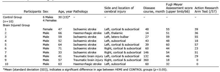
This pilot case-controlled prospective observational study included 10 adults with brain injury (HEMI) and 10 control participants (CONTROL) (see Table I for participants’ demographics). The inclusion criteria were: brain injury for at least 6 months caused by an acquired cerebral lesion (single stroke or traumatic brain injury); strength of paretic triceps brachii (rated at least at 3/5 on the Held-Deseilligny Scale, corresponding to extension of the forearm against slight resistance); and no anti-spastic treatment during the 3 previous months. Exclusion criteria were: severe cognitive disorders with limited comprehension of basic instructions; neurodegenerative conditions other than the acquired brain injury; elbow contracture (loss of passive elbow extension or flexion); and upper limb pain during movement. Ethics approval was obtained from the local institutional review board at Paul Sabatier University Hospital (No. 07-0716, Toulouse, France) and written informed consent was obtained from all participants. The study was conducted in accordance with the amended Declaration of Helsinki and conforms to all STROBE guidelines, reporting the required information accordingly.
Net torque around the elbow joint was recorded at 1 kHz using a Con-Trex MJ calibrated dynamometer (CMV AG, Dubendorf, Switzerland).
The surface electromyographic signal (EMG) was recorded at 1 kHz using Ag–AgCl bipolar electrodes in bipolar configuration with an inter-electrode distance of 20 mm,, using an MP150 system (Biopac Systems Inc., Goleta, CA, USA). The reference electrode was placed on the left ulnar head. Biceps brachii and brachioradialis were selected as the elbow flexors acting as antagonist muscles during elbow extension.
Recordings were made on the non-dominant side in the CONTROL group and on the paretic side in HEMI group.
Torque and EMG data were synchronized automatically using a rectangular triggering pulse signal and analysed offline.
The experimental procedure comprised 2 steps.
First step. To perform the following clinical assessment (7): spasticity of elbow flexors, limitation to active elbow extension, motor selectivity and motor function were assessed respectively, using the Tardieu scale, a goniometer during repetitive and dynamic voluntary elbow extensions at a preferred rate, the upper limb motor section of the Fugl-Meyer Assessment scale, and the Action Research Arm Test (Table I). Maximal net elbow extension torque value was taken as a functional marker of triceps brachii paresis, the Fugl-Meyer score as a motor selectivity assessment, and the Action Research Arm Test as a motor function assessment.
Table I. Participants’ demographics
Second step. Participants were seated on the dynamometer chair with their upper body strapped firmly, the upper arm positioned along the trunk, and the elbow flexed at 90° (Fig. 1). The participants exerted 3 isometric maximal voluntary contractions of the elbow in flexion and in extension for a duration of 5 s, with 1 min rest between each contraction and 3 min rest between flexion and extension contractions. After collection of maximal voluntary contraction data, participants performed 2 sets of 5-s elbow isometric extension sub-maximal contractions while receiving visual feedback on their actual torque in relation to a target torque. Each set included 6 contractions at 25% Maximal Voluntary Contraction (MVC), corresponding to a level of force required for daily activities (8). The time between each contraction was 30 s; each set was separated by a 3-min rest period to minimize fatigue.
Fig. 1. Illustration of the arm and forearm positions used to perform torque and electromyographic recordings during isometric elbow extension on the calibrated dynamometer.
Net torque was low-pass filtered at 100 Hz with a 6th-order zero-lag Butterworth filter. EMG data were 10–400-Hz band-pass filtered (4th-order zero-lag Butterworth filter), full-wave rectified, and smoothed at 9 Hz to obtain the linear envelopes. The co-contraction index was determined as the ratio (expressed in percentage) between the root mean square value of the elbow flexors EMG envelopes during the sub-maximal elbow extensions and the root mean square of the same muscle during the highest maximal voluntary elbow flexion contraction (9).
Non-parametric analysis using Wilcoxon rank-sum test was performed to compare the co-contraction index and the maximal net elbow extension torque between HEMI and CONTROL groups.
Non-parametric Spearman’s correlations (rs) were performed to investigate the relationship between co-contraction index, elbow-flexor spasticity, maximal net elbow extension torque with: (i) limitation to active elbow extension, (ii) elbow-flexor spasticity, (iii) Fugl-Meyer Assessment score for the upper limb, and (iiii) Action Research Arm Test. It is notable that that there was a significant difference in age between the HEMI and CONTROL groups (Table I). However, preliminary analysis of the data showed a lack of any correlation with age (p > 0.05), enabling the results to be interpreted independently of age.
A higher co-contraction index occurred in the HEMI group compared with the CONTROL group (w19 = 3.4, p < 0.01), with a mean difference (SD) of 7.6±12.9%. Lower net elbow extension torque during maximal voluntary contraction was found in the HEMI compared with the CONTROL group (w19 = –2.34, p<0.05), with a mean difference (SD) of 17.6 ± 17 Nm. Results of the Spearman’s correlations are shown in Table III.
This pilot study aimed to elucidate the consequences of spasticity and spastic co-contraction of elbow flexors on limitation of active elbow extension in adults with brain injury. A strong association was found between the co-contraction index and (i) the limitation of active elbow extension, (ii) the Fugl-Meyer Assessment score, and (iii) the score on Action Research Arm Test. Conversely, no significant correlation was found between spasticity and any of the variables cited above.
These results are thus the first to show that spastic co-contraction primarily contributes to a deficit in active elbow extension in adults with brain injury, which occurs even in the absence of spasticity (see, for example, Table II, HEMI participants 5 and 10). These findings confirm the absence of an association between spasticity and spastic co-contraction, supporting the idea that they refer to different forms of overactivity with different underlying physiological mechanisms (1). Most importantly, in agreement with a suggestion made in a previous report (10), these results unequivocally establish that spasticity and spastic co-contraction have different functional repercussions with regards to impaired motor function in adults with brain injury.
It is well known that selective muscle activation is necessary for skilled and coordinated upper limb movements, which implies concomitant activation of agonists and relaxation of antagonists. Lesions that damage the corticospinal pathways, such as stroke or traumatic brain injury, cause long-lasting impairment of the ability to produce selective patterns of EMG activity (11). However, it has been shown that, during rehabilitation, the decrease in co-contraction index is correlated with the improvement in Fugl-Meyer score among post-stroke subjects (12). In agreement with a previous study (4), our finding of a strong association between the co-contraction index and Fugl-Meyer Assessment score thus highlights the importance of impaired motor selectivity as a mechanism that contributes to greater spastic co-contraction.
The adverse consequence of the presence of spastic co-contraction on upper limb motor impairment is further supported by the significant association between the co-contraction index and the score on Action Research Arm Test, taken to reflect upper limb functional limitation. Furthermore, by analysing EMG-based assessment of spastic co-contraction in patients with a wide range of motor impairment, our results highlight that the more severe the motor impairment, the greater the co-contraction index.
These results also failed to show an association between the net elbow extension torque during maximal voluntary contraction, taken as a functional marker of triceps brachii paresis, and either the limitation of active elbow extension, the score of Fugl-Meyer Assessment or of the Action Research Arm Test. These findings indicate that motor weakness is not a primary factor limiting active elbow extension, and lead to the conclusion that non-selective motricity and function impairment are not directly linked to motor weakness.
Taken together, the above results demonstrate the detrimental impact of spastic co-contraction on upper limb motor function, leading to abnormal restricting arm movement patterns, especially in subjects with more severe motor impairment.
Despite strong evidence of improvement in spasticity induced by botulinum toxin treatment, few studies have shown effectiveness in improving active function and active movement (13, 14). A limitation that may explain this lack of efficacy in active function is the use of spasticity, as assessed during passive stretching, as a marker of muscle overactivity during movement (15). In contrast, the change in spastic co-contraction after injection of botulinum toxin has been poorly studied. From a clinical perspective, these results are in line with the Subcommittee of the American Academy of Neurology (13), which recommend the use and development of a method and outcome regarding motor function and active movement. We support the requirement to report active range of motion as an outcome of muscle overactivity treatments (7), and the use of EMG-based quantification of spastic co-contraction as a relevant tool for providing effective interventions related to altered recruitment of antagonist muscles and limitation of active movement.
Table II. Co-contraction index, net elbow extension torque during maximal voluntary contraction and clinical characteristics
Table III. Spearman correlations (95% confidence interval) for HEMI participants between co-contraction index, net elbow extension torque during maximal voluntary contraction and clinical variables
Although a limitation of this pilot study is the small sample size, significant findings were made relative to the link between spastic co-contraction and clinical scores. Any generalization of these results, however, should be viewed with caution, especially because hemiparetic subjects had different aetiologies of brain injury (i.e. stroke and traumatic brain injury) and that little is known about the influence of the type of brain injury on spastic co-contraction.
This pilot study assessed the co-contraction index during submaximal isometric contraction, while spastic co-contraction is sensitive to stretch (1). Thus, future research should investigate co-contraction during active elbow extension.
The results of this pilot study suggest that spastic co-contraction alters upper limb function in subjects with hemiparetic brain injury. These findings may partially explain the lack of data concerning the efficacy of treatments, such as botulinum toxin to improve upper limb function (6, 13, 14). Although measurement of spasticity is usually performed to assess muscle overactivity, these results highlight the importance of considering spastic co-contraction to assess active motor function, and support further studies on changes in spastic co-contraction after injection of botulinum toxin, in connection with the improvement in active function. Practical applications arising from this work are to improve the assessment of factors that restrict movement and implementation of treatments aimed at improving motor function in subjects with brain injury.
Alexandre Chalard is an employee of Ipsen Innovation within the framework of a Conventions Industrielles de Formation par la REcherche (CIFRE) PhD fellowship. Others authors in this study declare that they have no conflict of interest in relation to this study.


 Click to show fullsize
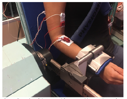 Click to show fullsize
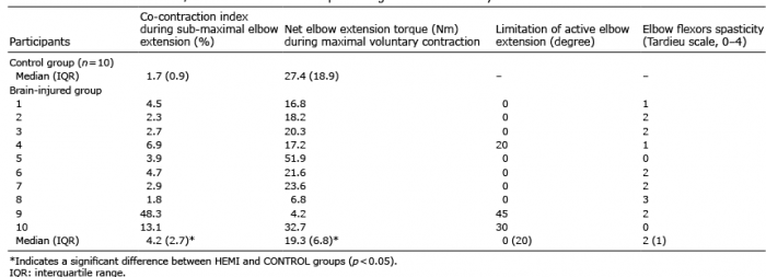 Click to show fullsize
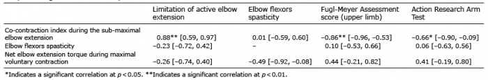 Click to show fullsize