Niina Hieta1 and Eija Hiltunen-Back2
Departments of Dermatology, 1Turku University Hospital and University of Turku, FIN-20520 Turku and 2University of Helsinki and Helsinki University Central Hospital, Helsinki, Finland. E-mail: niina.hieta@utu.fi
Accepted Mar 24, 2016; Epub ahead of print Mar 30, 2016
Recurrent vulvar ulceration is usually caused by herpes simplex virus (HSV) type 1 and 2 infections. Differential diagnosis includes erosive lichen planus, lichen sclerosus, mucous membrane pemphigoid, pemphigus, and Behçet’s disease. Lipschütz acute vulvar ulcer, occurring in adolescent women, is a non-recurring, rare ulcer associated with an immunologic reaction to a distant source of infection or inflammation (1). We report here the first case of human herpes virus 7 (HHV-7) in recurrent vulvar ulceration.
CASE REPORT
A 24-year-old woman presented with repeated vulval ulceration since the age of 15 years. The symptoms usually occurred less than once a year, always after an upper respiratory tract infection. One or more dark spots appeared on labia minora and enlarged, forming a deep, extremely painful ulcer. HSV cultures were repeatedly negative. Empirically given acyclovir and valacyclovir did not help.
One week before visiting the clinic, the patient had had sore throat, headache, myalgia, fever, vomiting, and cough. A few days later she had again noticed an ulcer. At the inner side of the left labia minor a 1 cm ulcer was seen, with erythematous edges under the fibrinous layer. The histopathological diagnosis from the erythematous area was a unspecific benign ulcer. Some oedema, capillaries and a large number of neutrophils were seen, but no vasculitic changes. The white blood cell count was slightly elevated (8.5 × 10⁹/ml; normal 3.4–8.2 × 10⁹/ml) as was C-reactive protein (18 mg/l; normal < 10 mg/l). Serological tests were negative for syphilis, HIV, cytomegalovirus (CMV) and parvovirus. Primary infection of Epstein Barr virus (EBV) or Chlamydia pneumoniae was not detected based on the levels of antibodies
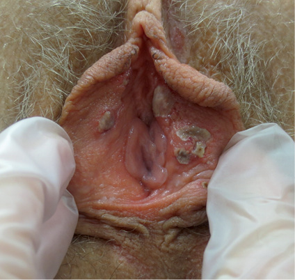
The next time the lesion appeared, the patient began treatment with prednisolone, 30 mg. The pain was clearly milder and the ulcer smaller (Fig. 1). Biopsy from the acute ulcer was studied using PCR for human herpes virus group (CMV, EBV, HSV-1 and -2, VZV, HHV-6, and HHV-7) and for Mycoplasma pneumoniae. The test was positive for HHV-7, and negative for other viruses and mycoplasma. The ulcer healed well, leaving a slightly paler area. Four months later, a biopsy was taken from the healthy mucosa in the area of the previous ulcer. The PCR test was negative for the human herpes virus group including HHV-7. Following a symptom-free period the patient again had a respiratory viral infection followed by vulvar ulcer. A biopsy from the ulcer was positive for HHV-7, but negative for other human herpes viruses. The patient has been using naproxen for pain and fever, when necessary, ever since childhood. She has also taken naproxen during the last years without the appearance vulvar symptoms.
Fig. 1. Fibrinotic, erythematous ulcers on the inner aspects of the labia minora 5 days after the patient had started with prednisolone.

The patient was positive for HLA-B*51:01, as are approximately 10% of the Finnish population. Behçet’s disease was ruled out, because the patient was found to have no ocular changes at the ophthalmologist’s consultation, no other skin changes, no vascular or neurological symptoms, and the pathergy test was negative. The patient had had constant oral aphthae-like ulcers, but their appearance reduced to approximately once in 2 months after changing her regular toothpaste to toothpaste without lauryl sulphate. A swab sample from an oral ulcer was negative for HHV-7 and other human herpes viruses, studied using PCR, as for samples from the vulvar ulcer.
DISCUSSION
HHV-6 and HHV-7 are closely related members of the human herpes virus group. Together with cytomegalovirus they belong to the sub-family of β-herpesviruses (2). Aciclovir, valaciclovir, and famciclovir, which are effective for HSV infection, have no effect on HHV-7. HHV-6 and HHV-7 are both acquired in early childhood. Exanthema subitum, usually caused by HHV-6, may also be a rare clinical presentation of primary HHV-7 infection. After infection in infancy, salivary glands continue producing HHV-7. In immunocompromised patients, HHV-7 may cause acute febrile respiratory disease, fever, rash, vomiting, diarrhoea, and encephalomyelitis (3).
Apart from exanthema subitum, HHV-7 rarely causes dermatological symptoms (2). A patient with asymmetric periflexural exanthema associated with HHV-7 infection has been described (4). In children with fever and maculopapular rash, HHV-7 was detected in 9/100 patients aged 2 months to 14 years (5). In addition, drug eruption with eosinophilia and systemic syndrome (DRESS) has been associated with reactivation of HHV-7, either alone or together with HHV-6 (6, 7). There is also some evidence on the role of HHV-7 in lichen planus (8). The role of HHV-6 and HHV-7 in pityriasis rosea is under debate (2, 9).
Kikuchi-Fujimoto disease (KFD), also known as histiocytic necrotizing lymphadenitis, is a self-limited febrile illness primarily of female patients under 30 years of age, often accompanied by chills, myalgia, sore throat, skin rash, and cervical lymphadenopathy. HHV-7 was found in 3/30 patients with KFD (10). Specific DNA from HHV-7 has been found in the lymph nodes in a 24-year-old female patient with KFD (11).
The prevalence of Behçet’s syndrome varies in different populations, being extremely rare in Finland. Interestingly, a Dutch group found HHV-7 DNA in cerebrospinal fluid of a 27-year-old female patient with Behçet’s syndrome (12). Idiopathic vulvar aphthosis with no infectious or autoimmune aetiology has been described in non-sexually active young girls (13), causing both pain and emotional distress. However, HHV-7 was not searched for in these studies (13, 14). Treatment consists mainly of supportive care and symptom relief. Local and systemic corticosteroids may be helpful, as for our patient.
To our knowledge, this is the first report of HHV-7 found in recurrent vulvar ulcer, but not in the non-lesional vulvar skin during the symptom-free period. We therefore suggest that HHV-7 may be involved in the pathogenesis of vulvar ulcers, but larger studies on more patients are needed.
REFERENCES
